Jonathan A. Sherratt, Department of Mathematics, Heriot-Watt University
Mathematical Modelling of Scar Tissue Formation
Biological Background
 The initial response to injury is bleeding and the formation of a
blood clot. The upper portion of the clot dries out to form the scab,
while the lower part is the setting for many of the key processes of
wound healing. In particular, cells known as fibroblasts move into the
blood clot from surrounding tissue, breaking it down and replacing it
with scar tissue. This is composed of the same main protein (collagen)
as normal skin, but with differences in details of composition. Most
crucially, the protein fibres in normal tissue have a random
(basketweave) appearance, while those in scar tissue have pronounced
alignment in a single direction.
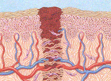
The figure shows a schematic illustration of a wound as it is healing.
The two layers of the skin, epidermis and dermis, are indicated, with
the blood clot between them. Fibroblast cells are illustrated,
migrating into the blood clot.
A Discrete-Cell Model
 John Dallon, Philip Maini, Mark Ferguson and I developed a
discrete-cell model for the process of scar tissue formation.
We represent each cell in the wound area explicitly, with rules
governing the movement of cells, their division, and their interaction
with surrounding protein fibres. The key behaviour is that the cells
tend to move in the direction in which nearby fibres are oriented;
also, the protein fibres are reoriented by the cells, towards their
direction of movement.
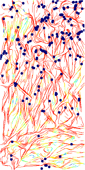
The figure shows a simulation of this model, showing the collagen fibre
network several days after injury. The black dots show the cells (only
10% of the cells are shown, for clarity). Note the pronounced alignment of
the collagen fibres, orthogonal to the plane of the skin.
The colour represents collagen density (red=high, blue=low).
Point to the image with the mouse to see the corresponding result when
an anti-scarring therapy is simulated. Our simulated therapy alters cell
behaviour in a way that mimics the effects of chemicals currently
being studied as possible anti-scarring agents.
Click on the image to load a movie of the model simulation.
A Continuum Model
 John Dallon and I have also developed continuum models for
collagen alignment by fibroblast cells. The model consists of
integrodifferential equations for fibroblast and collagen densities,
which are a function of space and orientation. The integral terms
enter the equations because of non-local interaction in orientation
space: a cell will interact with collagen fibres at the same point
even if the cell and fibre orientations are different.
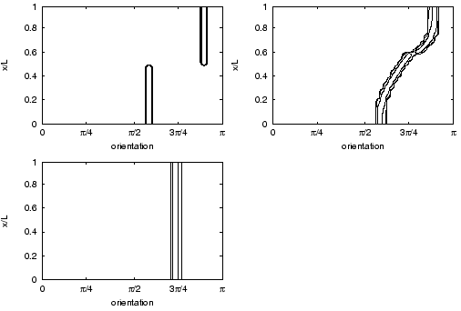
The figure shows a model simulation in which collagen fibres are
aligned with one uniform direction in one region of space (x), and another
direction elsewhere. The interaction between the collagen fibres and
the fibroblasts causes the fibres to be reorientated, over time, into
a single, intermediate direction.
The work described on this page is discussed in the following papers:
-
J. Dallon, J.A. Sherratt:
A mathematical model for fibroblast and collagen orientation.
Bull. Math. Biol. 60: 101-130 (1998).
Click to see the
 Full paper (PDF)
-
J. Dallon, J.A. Sherratt, P.K. Maini:
Mathematical modelling of extracellular matrix dynamics using discrete
cells: fibre orientation and tissue regeneration.
J. Theor. Biol. 199: 449-471 (1999).
Click to see the
 Full paper (PDF)
-
J. Dallon, J.A. Sherratt:
A mathematical model for spatially varying extracellular matrix alignment.
SIAM J. Appl. Math. 61 506-527 (2000).
Click to see the
 Full paper (PDF)
-
J. Dallon, J.A. Sherratt, P.K. Maini, M.W.J. Ferguson:
Biological implications of a discrete mathematical
model for collagen deposition and alignment
in wound repair.
IMA J. Math. Appl. Med. Biol.
17 379-393 (2000).
-
J.C. Dallon, J.A. Sherratt, P.K. Maini:
Modeling the effects of TGF-beta on extracellular matrix alignment in
dermal wound repair.
Wound Repair Regen. 9 278-286 (2001).
Click to see the
 Full paper (PDF)
Back to Jonathan Sherratt's Research Interests
Back to Jonathan Sherratt's Home Page
 John Dallon, Philip Maini, Mark Ferguson and I developed a
discrete-cell model for the process of scar tissue formation.
We represent each cell in the wound area explicitly, with rules
governing the movement of cells, their division, and their interaction
with surrounding protein fibres. The key behaviour is that the cells
tend to move in the direction in which nearby fibres are oriented;
also, the protein fibres are reoriented by the cells, towards their
direction of movement.
 The initial response to injury is bleeding and the formation of a
blood clot. The upper portion of the clot dries out to form the scab,
while the lower part is the setting for many of the key processes of
wound healing. In particular, cells known as fibroblasts move into the
blood clot from surrounding tissue, breaking it down and replacing it
with scar tissue. This is composed of the same main protein (collagen)
as normal skin, but with differences in details of composition. Most
crucially, the protein fibres in normal tissue have a random
(basketweave) appearance, while those in scar tissue have pronounced
alignment in a single direction.
 John Dallon and I have also developed continuum models for
collagen alignment by fibroblast cells. The model consists of
integrodifferential equations for fibroblast and collagen densities,
which are a function of space and orientation. The integral terms
enter the equations because of non-local interaction in orientation
space: a cell will interact with collagen fibres at the same point
even if the cell and fibre orientations are different.
 Full paper (PDF)